 home
home
Standard histological staining
The morphological structure of tissue can be depicted with high accuracy by means of histological sections. Staining results depend on numerous factors, includi…


en


Menu
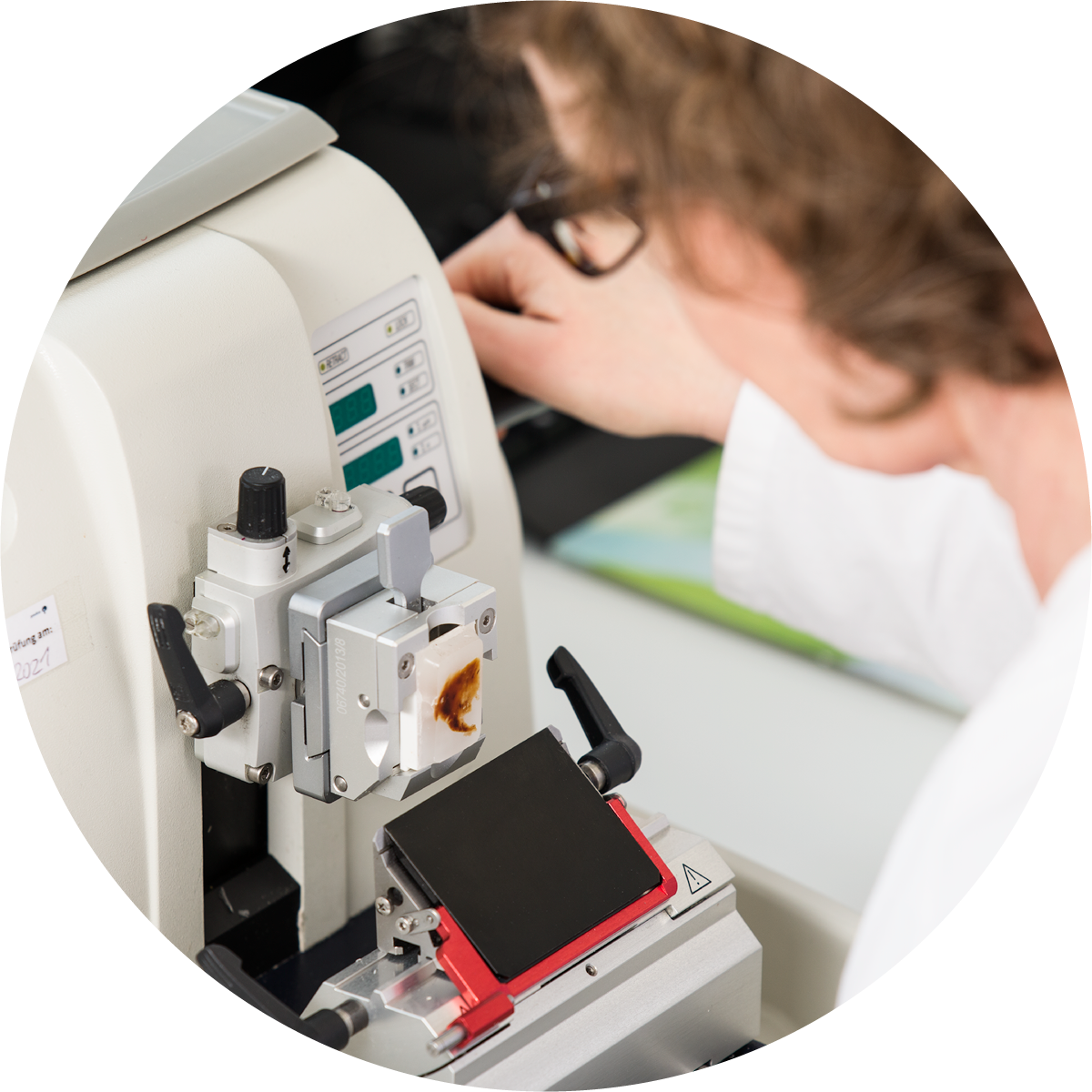
The morphological structure of tissue can be depicted with high accuracy by means of histological sections. Staining results depend on numerous factors, including pH of the buffer solution, staining time and method of fixation. Haematoxylin, for example, is used to stain cell and tissue structures, such as cell nuclei, mitochondria, myelin, elastin and collagen fibres. Additional information may be obtained from counterstaining (differential staining), using a dyestuff in high contrast to hematoxylin stain. Counterstaining, using eosin, is a classical approach for which cationic structures are stained (e.g. proteins).
Identification of antisera, immunoglobulin fractions and monoclonal antibodies to a growing number of clinically relevant tissue antigens has led to an enormous enlargement of immunohistochemical analyses in both quality and quantity. Antibody titre and dilution as well as incubation time and temperature are closely linked to each other with regard to their influence on the quality of immunohistochemical staining. These factors are varied either separately of each other or each of them in itself to achieve clearly recordable differences in staining quality. Parameters are varied primarily for the purpose of accomplishing staining of optimum specificity against minimum background.
The morphological structure of tissue can be depicted with high accuracy by means of histological sections. Staining results depend on numerous factors, includi…
Read more

 Catalogue
We recommend to use the Adobe Acrobat Reader for the best experience.
Copyright: 2020 provitro AG,
Charitéplatz 1, 10117 Berlin
provitro AG
Charitéplatz 1, 10117 Berlin
Tel:
+49.30.450 578 358
Email:
sales@provitro.com

Provitro stands for more than ten years of experience in commercialisation of cell culture technologies and tissue microarrays backed up by scientific knowledge and resources of immunohistochemical analyses at the Pathological Institute of Charité Universitätsmedizin Berlin. We have added medical research to our own know-how in biotechnology to create solutions to common problems in life sciences. Provitro’s team has raised its sights to the interface between research and development to hammer out brand-new solutions to the benefit of future research projects. These are the hallmarks: competence, vision and sustainability.